CT vs. MRI
Computed Tomography (CT)
Computed tomography (CT) is a modern imaging tool that combines X-rays with computer technology to produce a more detailed, cross-sectional image of your body. A CT scan lets your doctor see the size, shape, and position of structures that are deep inside your body, such as organs, tissues, or tumors.
Magnetic Resonance Imaging (MRI)
Magnetic resonance imaging (MRI) is another modern diagnostic imaging technique that produces cross-sectional images of your body. Unlike CT scans, MRI works without radiation. The MRI uses magnetic fields and a sophisticated computer to take high-resolution pictures of your bones and soft tissues.
The table below outlines which image modality MRI or CT may be considered depending on what your physician is looking for.
Head / Brain
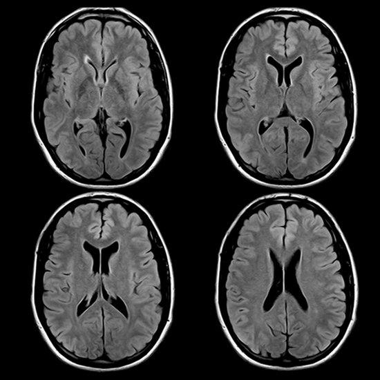
MRI
Arteriovenous malformations
Encephalomalacia
Epilepsy and adult onset seizures
Hemangiomas
Hydrocephalus
Infection / Inflammation
Infratentorial tumors
Lacunar infarcts
Lyme disease
Meningiomas
Multiple sclerosis
Orbit and optic nerve disease
Pituitary dysfunction
Posterior fossa abnormalities (acoustic neuroma and skull base pathology)
Space occupying lesions
Stroke—(Use diffusion-weighted MR imaging)
Supratentorial tumors
Syringomyelia and Chiari malformations
Vascular/congenital abnormalities
White matter disease
CT
Acute trauma
Calcified lesions
Suspected acute intracerebral hemorrhage
Suspected subarachnoid hemorrhage (“The worse headache of my life.”)
ENT

MRI
Disease of the larynx (staging)
Paranasal sinus diseases (soft tissue masses, CA staging)
TMJ meniscal and soft tissue evaluations
Sensorineural hearing loss
CT
Cholesteatoma
Conductive hearing loss
Evaluation of the oro-, hypo-, and nasopharynx
Facial bone trauma
Orbital trauma
Ossicular or vestibulocochlear deformities
Otosclerosis/otospongiosis continuum
Sinusitis
Salivary gland disease
TMJ – bone destruction or arthritis
Spine
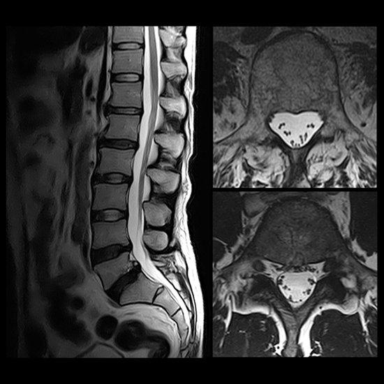
MRI
Any intrinsic disease of the cord
Infection / inflammation
Disc disease (cervical, thoracic, lumbar)
Multiple sclerosis and demyelinating disease
Myelopathy – all levels
Paraspinal masses
Postoperative evaluation (differentiate disc herniation from scar tissue)
Scoliosis (MRI best to evaluate cord)
Spinal cord tumors
Syringomyelia, Chiari malformations, hydromyelia
Syrinx
Tethered cord
Vascular abnormalities
Vertebral column bone destruction by tumor (evaluation of spinal canal integrity only)
Vertebral osteomyelitis
CT
Post discography to assess the morphology of an intervertebral disc
Scoliosis (CT best for bone detail)
Vertebral fractures – all levels
Chest

MRI
Assessment of cardiac function
Brachial plexus and axillary pathology
Congenital heart lesions or cardiac abnormalities
Great vessel anomalies
Intracardiac or pericardial masses
Mediastinal masses
Vascular structures of the mediastinum and chest
Valvular disease
CT
Aortic dissection
Hilar and parenchymal nodules
Lung disease
Abdomen

MRI
Evaluation of renal vasculature (MRA)
MRCP recommended when
Diagnostic component needed before therapeutic ERCP
When ERCP is impossible
Patients who have failed ERCP
Patients who only require a diagnostic study
To exclude sclerosing cholangitis, pancreatic neoplasm or chronic pancreatitis
Tumor invasion of vena cava
CT
Abdominal aortic aneurysm
Benign and malignant disease of the liver
Diagnosis and staging of non-hepatic intra-abdominal infections and tumors
Screening examination for symptoms when detail of the liver, spleen, kidneys or pancreas is needed
Pelvis

MRI
Bladder carcinoma staging
Prostate carcinoma staging
Uterine carcinoma staging
Seminal vesical tumor invasion
CT
Nodal assessment
Pelvic Mass (useful, but ultrasound still preferred)
Pelvic pain
MUSCULOSKELETAL


MRI
Achilles tendon injury
Ankle – ligamentous injuries
Avascular necrosis
Bone and soft tissue tumors
Knee injury including meniscal, cartilaginous and tendinous
Metastatic disease of bone
Occult fractures not apparent on plain films
Osteoarthritis of hip, knee, shoulder and ankle
Osteochondritis dissecans
Osteomyelitis
Rotator cuff injuries
Tendon injuries of the elbow
CT
Complex fractures (3-D CT reconstruction)